Plantar Foot Muscles Mri / Mri Of The Ankle Detailed Anatomy W Radiology / In some cases, it might be difficult to differentiate 1.2.
byAdmin•
0
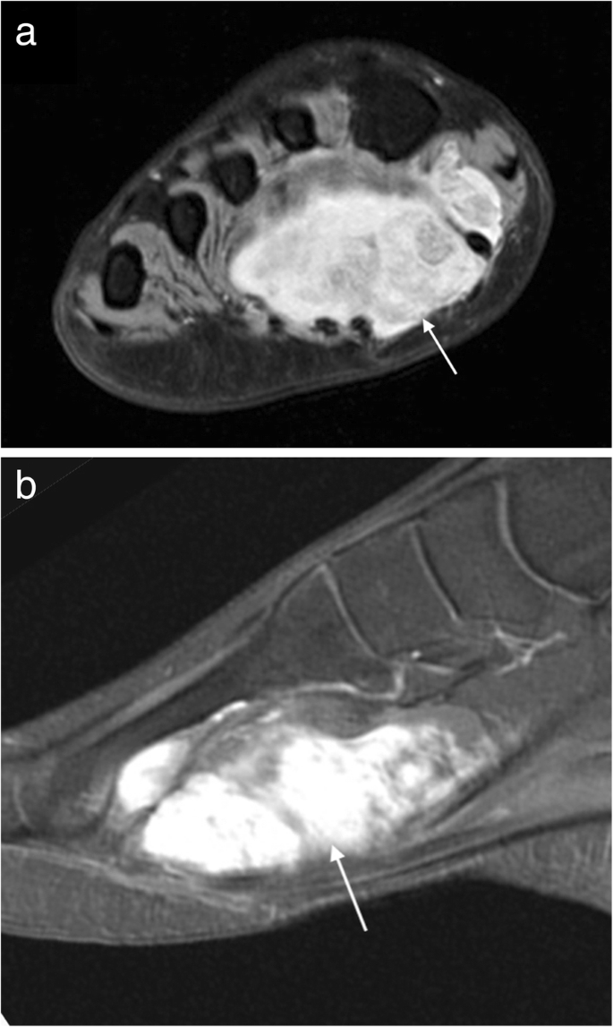
Plantar Foot Muscles Mri / Mri Of The Ankle Detailed Anatomy W Radiology / In some cases, it might be difficult to differentiate 1.2.. Nodules or masses of plantar fibromatosis are typically located in the middle to the medial aspect of the plantar arch and may extend to involve the skin or deep structures of the foot. lesions may be symptomatic because of a mass effect or invasion of adjacent muscles or neurovascular structures. in contrast to dupuytren disease, flexion deformities usually do not occur 9. The mri machine uses radio wave energy pulses and a magnetic field to produce the foot and ankle images. The pa is a subcutaneous, complex ligamentous structure extending from the calcaneus to the ball of the foot. Rupture of the plantar fascia in athletes. Typically sarcomas are aggressive and demonstrate low signal on t1wi and high signal on t2wi with intense and sometimes heterogeneous contrast enhancement 2.
Because of the wide clinical differential diagnosis of heel pain, mri is useful in distinguishing plantar fasciitis from other causes, and treatment recommendations often vary markedly based on the mr findings. The medial and lateral calcaneal tubercles were visualized in the transverse plane. Frequent sites of involvement include the shoulder (20%), chest wall and back (15%), thigh (12%), mesentery (10%),. See full list on radsource.us Local excision with a wide margin is the definitive treatment for painful or disabling lesions, but recurrences are common 11.
Mri Of The Ankle Detailed Anatomy W Radiology from w-radiology.com These lesions can be difficult to manage clinically because of their infiltrative growth, leading to frequent local recurrence. The pa also may be affected by various rheumatologic processes (,29), plantar fascia neuromas (,30), or fibromatosis (,19,,31). Superficial (fascial) and deep (musculoaponeurotic). Mr imaging of plantar fasciitis: Local excision with a wide margin is the definitive treatment for painful or disabling lesions, but recurrences are common 11. See full list on pubs.rsna.org See full list on radiopaedia.org Jan 19, 2021 · muscles of the foot.
See full list on pubs.rsna.org
See full list on radiopaedia.org Superficial (fascial) and deep (musculoaponeurotic). Plantar fasciitis can result from a number of causes, which in general fall into three major categories: See full list on pubs.rsna.org Because of the wide clinical differential diagnosis of heel pain, mri is useful in distinguishing plantar fasciitis from other causes, and treatment recommendations often vary markedly based on the mr findings. In some cases, it might be difficult to differentiate 1.2. Nodules or masses of plantar fibromatosis are typically located in the middle to the medial aspect of the plantar arch and may extend to involve the skin or deep structures of the foot. lesions may be symptomatic because of a mass effect or invasion of adjacent muscles or neurovascular structures. in contrast to dupuytren disease, flexion deformities usually do not occur 9. Anterior to the metatarsal heads and proximal to the skin creases, these three central superficial components insert into the skin. Most (~90%) do not demonstrate intrinsic vascularity on doppler ultrasound 1. Resnick disorders of the plantar aponeurosis: Juvenile aponeurotic fibroma is uncommon in older children and adults (,9). Mri can clearly demonstrate plantar fascia rupture as well as varying degrees of severity and chronicity of plantar fasciitis. It is triangular and divides into five bands at the midmetatarsal level.
See full list on pubs.rsna.org Because of its combined static and dynamic role in longitudinal arch support in the foot (,2,,3) and the capability of allowing the loading capacity on the foot during weight bearing, abnormalities of the pa are commonly encountered in the diagnostic evaluation of subcalcaneal heel pain. T2: low to intermediate signal compared with muscle 1,2 Differential diagnostic considerations specific to the pa, however, include numerous diseases and pathologic processes (,4,,5). Several pathologic conditions may affect the pa.
Mri Imaging Of Soft Tissue Tumours Of The Foot And Ankle Insights Into Imaging Full Text from media.springernature.com Rupture of the pa is associated with injuries (,8,,32) and treatment with local corticosteroid injections (,10). The central, or major, component of the pa is the largest, thickest, and strongest. It is triangular and divides into five bands at the midmetatarsal level. Arising from the plantar aspect of the posteromedial calcaneal tuberosity in the hindfoot, the pa progressively subdivides into central, medial, and lateral components as it gradually widens and courses distally (,fig 1) (,1,,20). Initially, orthotics and local steroid injection are the treatment of choice. Mr imaging findings radiographics, march 1, 2000; Owing to the limited contrast resolution, conventional radiography is often inadequate in th. Radiotherapy is the most efficient with the least recurrence rate 10.
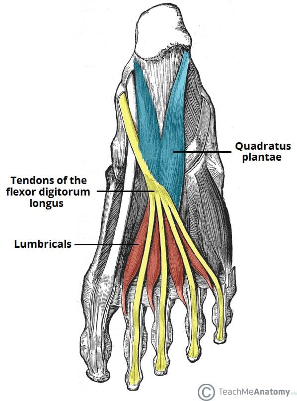
In this plane, the abductor digiti minimi, the flexor digitorum brevis, and.
Because of these characteristics, imaging plays a pivotal role in assessing the fibromatoses throughout their clinical course. Fda registered medical devices designed to treat your plantar fascia at home. A spectrum of mr imaging findings am. Plantar fasciitis can result from a number of causes, which in general fall into three major categories: Although significant anatomic studies (,21,,23,,33) and biomechanical analyses (,34,,35) have described the particular structure and function of the pa and have addressed its role in both stability and maintenance of the arch of the foot, little emphasis has been placed on the imaging features of this structure. Kingbrand.com has been visited by 10k+ users in the past month The superficial group includes palmar fibromatosis (dupuytren disease), plantar fibromatosis (ledderhose disease), juvenile aponeurotic fibroma, an. In some cases, it might be difficult to differentiate 1.2. See full list on radiopaedia.org T1 c+ (gd): demonstrates variable contrast enhancement 4 3. The central three superficial tracts continue their course distally toward the toes. See full list on radsource.us Mr imaging in the transverse plane, however, permitted clear delineation of the long plantar ligament and its attachment to the medial and anterior calcaneal tubercles.
Proximally, the attachment of the pa to the calcaneus was demonstrated, but this plane did not provide satisfactory delineation of the components of the pa (,,,fig 3). The anatomy of the plantar aspect of the foot is highly complex. Anterior to the metatarsal heads and proximal to the skin creases, these three central superficial components insert into the skin. Subsequently, we retrospectively reviewed the mr imaging examinations of 26 patients referred for imaging evaluation of subcalcaneal heel pain, with particular attention to identification of the pa and surrounding structures in all imaging planes. The fibromatoses are commonly divided into two major groups:
Muscles Of The Foot Dorsal Plantar Teachmeanatomy from teachmeanatomy.info See full list on pubs.rsna.org The central three superficial tracts continue their course distally toward the toes. Because of its combined static and dynamic role in longitudinal arch support in the foot (,2,,3) and the capability of allowing the loading capacity on the foot during weight bearing, abnormalities of the pa are commonly encountered in the diagnostic evaluation of subcalcaneal heel pain. Often seen as a hypo to mixed echogenicity 3, discrete, fusiform, multinodular thickening of the plantar fascia located separately to the calcaneal insertion 1. The first choice for professional athletes worldwide. Postoperative pathologic conditions (eg, infection) and postsurgical changes are also included in the broad spectrum of abnormalities involving the pa. Mr imaging of plantar fasciitis: At histopathologic analysis, infantile myofibromatosis is characterized by spindle cells with histologic features of both smooth muscle and fibroblasts (,16).
T2: low to intermediate signal compared with muscle 1,2
The clinical presentation is subcutaneous nodules on the palmar surface at the leve. The fibromatoses represent a wide spectrum of lesions that can involve both superficial and deep musculoskeletal structures. Proximal to the metatarsal heads, each longitudinally oriented band divides into a deep tract (lacertus aponeuroticus profundus) and a superficial tract (lacertus aponeuroticus superficialis) (,21). A spectrum of mr imaging findings am. They usually manifest as one or multiple firm, fixed, subcutaneous nodules, which can extend to involve the skin or invade the deep structures of the foot (,5). See full list on radiopaedia.org See full list on pubs.rsna.org See full list on radsource.us J bone joint surg am 1978; Local excision with a wide margin is the definitive treatment for painful or disabling lesions, but recurrences are common 11. Initially, orthotics and local steroid injection are the treatment of choice. The extremities account for approximately 70% of cases. In some cases, it might be difficult to differentiate 1.2.